Glymphatic System Dysfunction in Migraine: From Cortical Spreading Depression to Migraine Chronification
Article information

Abstract
Migraine has traditionally been understood as a neurovascular and neuroinflammatory disorder, but the glymphatic system, a brain-wide waste-clearance network mediated by cerebrospinal fluid and interstitial fluid exchange, has been proposed as a contributor to migraine pathophysiology. This review synthesizes current evidence on the role of glymphatic system dysfunction in migraine onset and chronification, integrating findings from preclinical animal models and multimodal clinical neuroimaging studies. Converging data suggest that cortical spreading depression during migraine attacks induces astrocyte swelling and aquaporin-4 (AQP4) depolarization, leading to physical collapse of the perivascular space, disruption of convective bulk flow, and impaired glymphatic clearance. This clearance failure may prolong the accumulation of nociceptive molecules, including calcitonin gene-related peptide, potassium ions, and glutamate, within the brain parenchyma, thereby exacerbating trigeminovascular sensitization and potentially contributing to migraine chronification. Clinically, alterations in neuroimaging markers such as the diffusion tensor imaging analysis along the perivascular space index and enlarged perivascular spaces have been associated with progressive fluid stagnation and an increased brain age gap in patients with chronic migraine. Glymphatic clearance failure may therefore contribute to the cycle of neuroinflammation and pain that sustains migraine. Future therapeutic strategies may benefit from moving beyond single-receptor blockade toward approaches that normalize the brain microenvironment, potentially by supporting glymphatic clearance through targeted pharmacological interventions and AQP4 channel modulation.
INTRODUCTION
The discovery of the glymphatic system, a specialized metabolic waste clearance pathway in the brain parenchyma, has significantly expanded the understanding of cerebrospinal fluid (CSF) circulation.1 According to classical circulation theory, CSF was primarily understood as a pressure-gradient-driven passive dilution pathway, produced in the choroid plexus, flowing through the ventricles and subarachnoid space, and draining into the venous sinuses via arachnoid granulations.1,2 However, this passive flow model proved insufficient to fully explain the active clearance of metabolic waste generated deep within the metabolically demanding brain parenchyma.1,2
Overcoming these limitations, in vivo two-photon imaging and fluorescent tracer studies3 demonstrated that a substantial portion of subarachnoid CSF penetrates the brain interstitial space and circulates throughout the parenchyma. This intricate system operates in three anatomical stages.1,4 First, subarachnoid CSF flows into the parenchyma along the peri-arterial Virchow-Robin space, driven by arterial pulsatility. Second, facilitated by aquaporin-4 (AQP4) water channels—highly expressed on astrocytic endfeet forming the outer boundary of the peri-arterial space—CSF rapidly enters the interstitial space and undergoes exchange with interstitial fluid (ISF) containing metabolic waste.1,5 Third, this fluid mixture, carrying metabolic byproducts and inflammatory mediators, is collected into the peri-venous space and ultimately drains via the meningeal lymphatic system into the deep cervical lymph nodes, connecting with the systemic immune system. The highly polarized localization of AQP4 on astrocytic endfeet is therefore a critical physiological feature that sustains this convective bulk flow and preserves central nervous system homeostasis.1,5
The discovery of this intrinsic brain waste clearance mechanism has opened a new perspective on the pathophysiological understanding of migraine, a complex neurovascular disorder. To date, the prevailing understanding of migraine pathophysiology has focused on the abnormal activation of the trigeminovascular system and the subsequent development of sterile neuroinflammation.6,7 According to this model, various neuropeptides released from trigeminal sensory nerve endings, such as calcitonin gene-related peptide (CGRP) and pituitary adenylate cyclase-activating polypeptide (PACAP), dilate meningeal blood vessels, degranulate mast cells, and promote the release of pro-inflammatory cytokines (e.g., tumor necrosis factor [TNF]-α, interleukin [IL]-1β), thereby transmitting and sustaining pain signals to the central nervous system.6,8 The clinical success of targeted monoclonal antibody therapies that block the CGRP signaling pathway has provided strong evidence for this established neuroinflammation and receptor-centric paradigm.8,9
However, this conventional paradigm has an important limitation: it largely overlooks how noxious molecules, once accumulated in local brain tissue and perivascular spaces (PVS) during a migraine attack, are ultimately processed and cleared.4 According to classical theories, inflammatory molecules should be passively diluted and eliminated via venous blood; however, this process alone cannot fully explain the frequent recurrence and chronification of migraine.
Recent evidence increasingly supports the hypothesis that dysfunction of the glymphatic system—the brain’s active waste clearance network—represents an important pathophysiological mechanism in migraine onset and chronification.10 By considering not only the generation of noxious substances through neuroinflammation but also their impaired clearance, an integrated pathophysiological cascade emerges: from inflammatory onset and clearance failure, to molecular accumulation, and ultimately to the induction and exacerbation of pain. A mechanistic analysis of glymphatic clearance failure helps address the limitations of current single-target therapeutic approaches.1 Based on this perspective, the present review examines the relationship between glymphatic dysfunction and migraine, integrating evidence from basic physiological research to clinical neuroimaging studies (Tables 1 and 2).
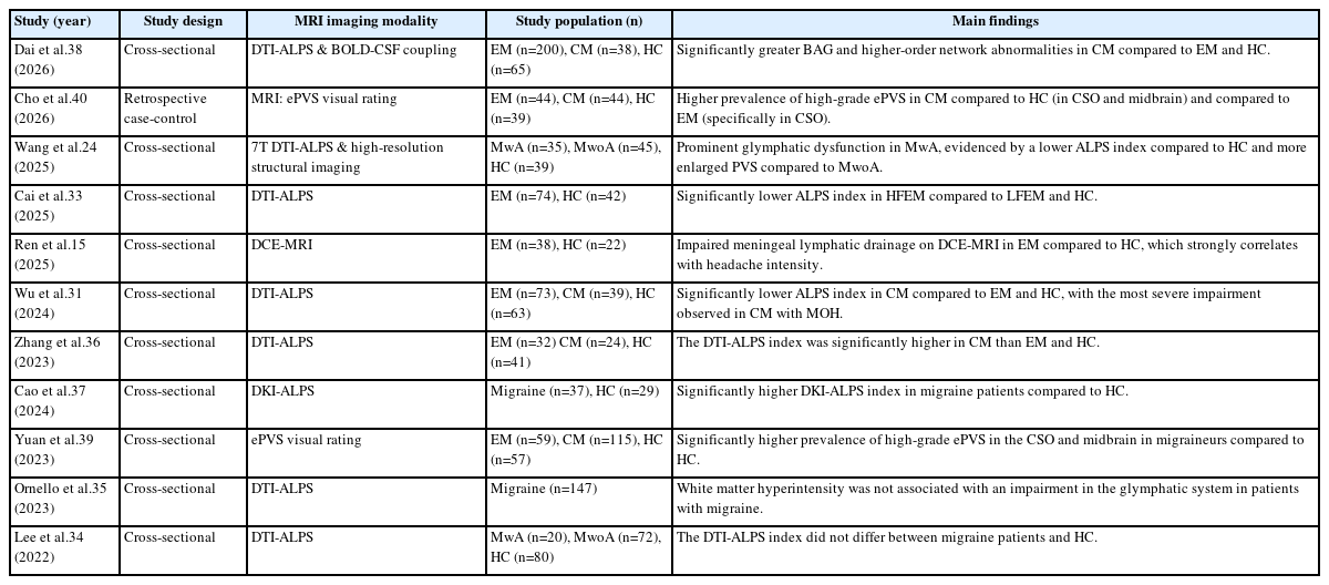
Clinical neuroimaging studies on glymphatic system dysfunction in migraine
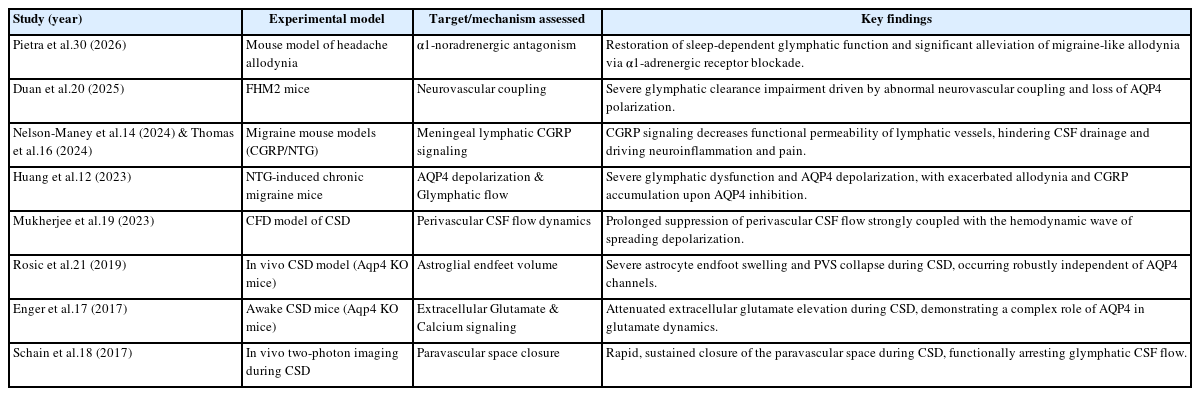
Preclinical and animal model studies on glymphatic impairment in migraine
METHODS
This article is a narrative review designed to synthesize current evidence on the role of the glymphatic system in migraine pathophysiology. While not a formal systematic review, a structured literature search was conducted to ensure a comprehensive overview of the topic. We searched the PubMed and Scopus databases for peer-reviewed articles published from January 2014 to July 2026, including advance online publications/e-pubs and preprints), focusing on recent advancements in both basic and clinical research. The search strategy utilized combinations of the following keywords: “migraine”, “glymphatic system”, “aquaporin-4”, “cortical spreading depression”, “perivascular space”, and “meningeal lymphatic”. We included original preclinical studies (e.g., in vivo animal models) and clinical neuroimaging studies (e.g., diffusion tensor imaging analysis along the perivascular space [DTI-ALPS], enlarged perivascular spaces [ePVS]) published in English. Studies focusing solely on non-migraine neurodegenerative diseases without relevance to headache disorders, non-peer-reviewed preprints (with the sole exception of one recent preprint providing critical preclinical insight into pharmacological restoration), and non-English articles were excluded. Following the initial screening of titles and abstracts, the full texts of relevant articles were reviewed and narratively synthesized.
PATHOPHYSIOLOGICAL MECHANISMS
1. Glymphatic dysfunction and activation of the trigeminovascular pain system
Under normal physiological conditions, key neuropeptides released during trigeminal nerve activation, such as CGRP and PACAP, are ultimately cleared into the meningeal lymphatic system or venous sinuses via the convective bulk flow of CSF and ISF (Figure 1A).4,11 However, when glymphatic clearance is impaired during an attack, these large-molecular-weight neuropeptides accumulate at elevated concentrations within the PVS and interstitial space.8 Accumulated CGRP continuously binds to CGRP receptor complexes located on the meningeal vascular walls, trigeminal ganglion (TG) neurons, satellite glial cells, and sensory nerve endings in the spinal trigeminal nucleus, inducing potent vasodilation and facilitating synaptic transmission.4 When CGRP receptor signaling within the TG is chronically activated, the gene expression of pro-inflammatory molecules and nociceptors is upregulated. This lowers the nociceptive threshold of local tissues, causing peripheral and central sensitization.8 Glymphatic clearance failure also enhances PACAP-mediated signaling, further contributing to the sensitization of the trigeminovascular system.11 Indeed, the finding that CGRP titers significantly increase and allodynia is exacerbated when migraine-model mice are administered TGN-020 (an AQP4 inhibitor) suggests that the glymphatic system serves as a major active clearance pathway regulating neuropeptide concentrations.4,12
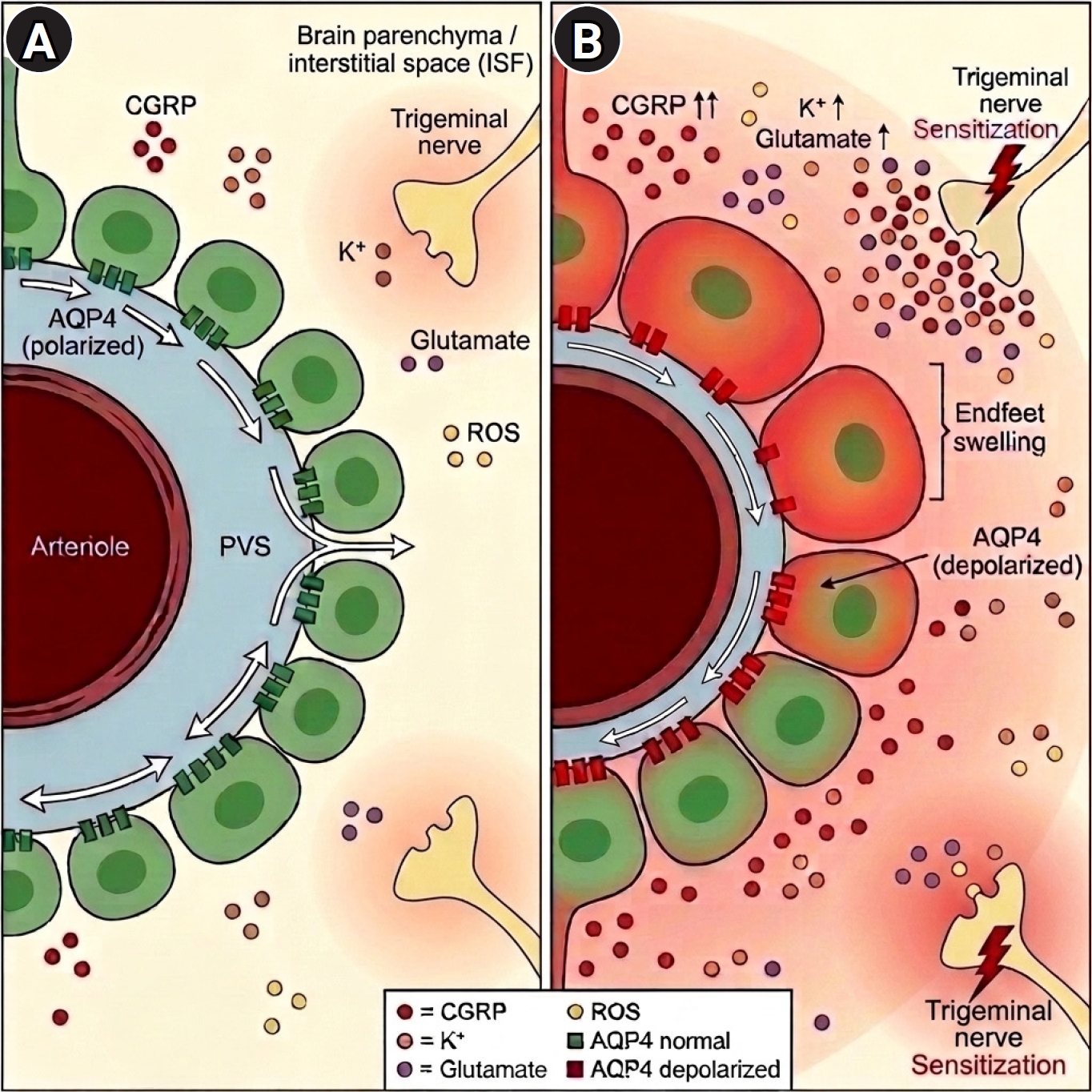
Glymphatic system impairment in migraine pathophysiology. (A) Normal: Cerebrospinal fluid (CSF) flows into the brain parenchyma along the arteriolar perivascular space (PVS), facilitated by polarized aquaporin-4 (AQP4) channels on astrocytic endfeet, depicted as green cells. CSF exchanges with interstitial fluid (ISF), supporting the clearance of metabolic waste from the brain. (B) Migraine: During or after a migraine attack, trigeminal neuroinflammation and cortical spreading depression may cause astrocyte swelling and AQP4 depolarization. These changes can collapse the PVS, reduce CSF inflow, and impair glymphatic clearance. As a result, nociceptive mediators, including calcitonin gene-related peptide (CGRP), potassium ions (K+), and glutamate, may accumulate around trigeminal nerve endings and promote trigeminal sensitization.
ROS, reactive oxygen species.
Alongside neuropeptide accumulation, reactive oxygen species (ROS) and pro-inflammatory cytokines (e.g., TNF-α, IL-1β) that remain in the microenvironment due to clearance failure markedly increase the excitability of pain transmission pathways.11 These accumulated, high-concentration inflammatory mediators substantially sensitize trigeminal sensory neurons, establishing a neuroinflammatory microenvironment that promotes attack induction and chronification.13 Sustained sterile neuroinflammation perpetuates pain symptoms, while elevated concentrations of ROS exacerbate oxidative stress, acting as a direct pathological factor that compromises neuronal membrane stability.4,6 Ultimately, this pro-inflammatory microenvironment and glial dysfunction further impair AQP4-mediated water transport, triggering a vicious cycle in which neuroinflammation leads to glymphatic decline, which in turn permits the accumulation of even more noxious substances (Figure 2).13
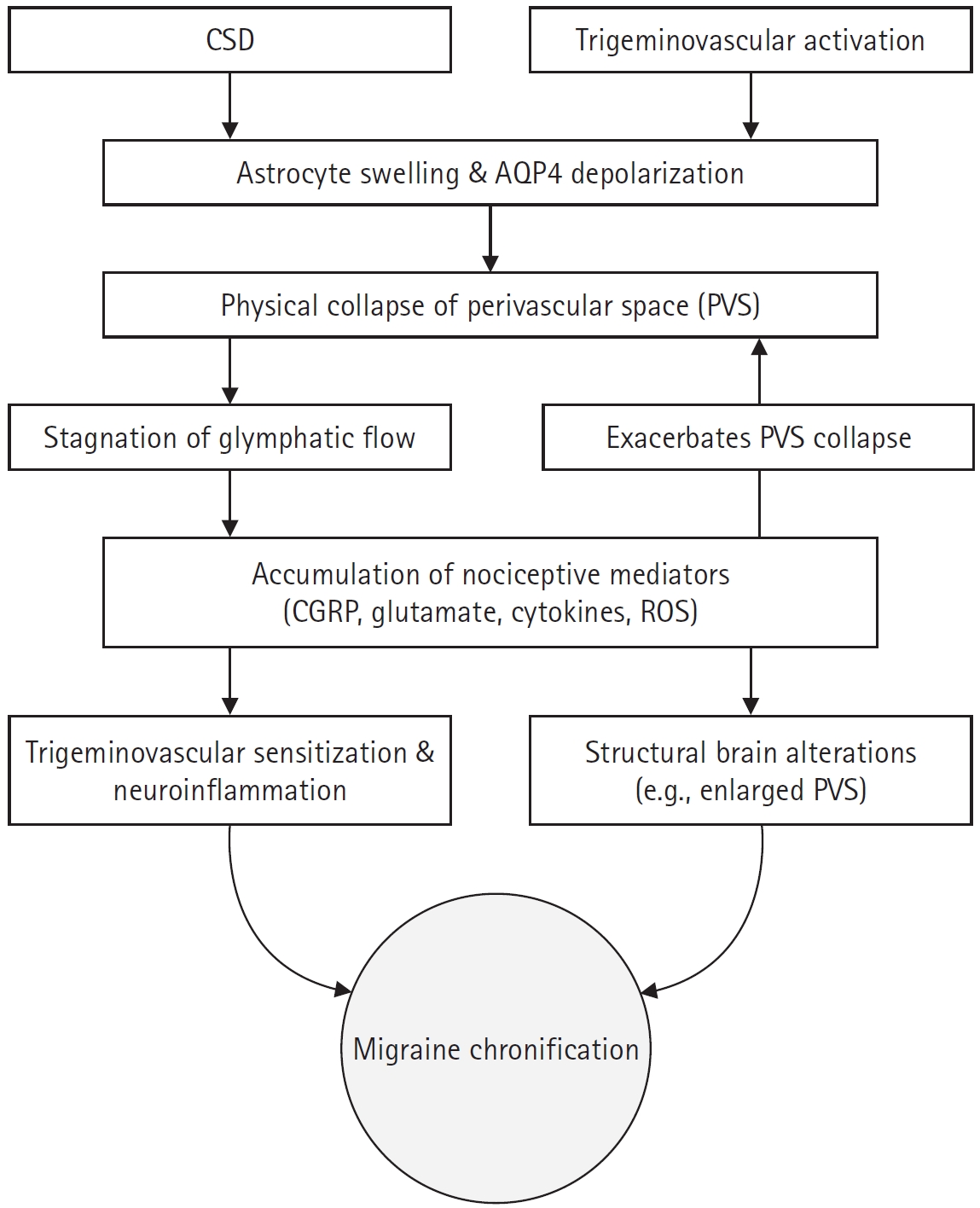
Mechanistic framework of migraine chronification driven by glymphatic dysfunction. Cortical spreading depression (CSD) and trigeminovascular activation are proposed to induce astrocyte swelling and aquaporin-4 (AQP4) depolarization within the brain microenvironment. These cellular changes may collapse the perivascular space (PVS), resulting in glymphatic flow stagnation. Subsequent clearance failure may allow nociceptive and excitatory molecules, such as calcitonin gene-related peptide (CGRP) and glutamate, to accumulate. These accumulated mediators may further activate the trigeminovascular system, establishing a self-reinforcing feedback loop that contributes to the transition to chronic migraine.
ROS, reactive oxygen species.
This decline in glymphatic clearance function is closely linked to the impairment of the meningeal lymphatic system, which serves as the final drainage pathway for CSF. Recent studies indicate a direct correlation between clearance impairment and headache intensity.14,15 Using 3T dynamic contrast-enhanced magnetic resonance imaging (MRI), Ren et al.15 demonstrated that patients with episodic migraine (EM) exhibited severe meningeal lymphatic clearance defects—such as significantly reduced contrast wash-in rates and delayed time-to-peak in the meningeal lymphatic vessels surrounding the superior sagittal sinus and sigmoid sinus—compared to healthy controls. Notably, these functional parameters of delayed meningeal lymphatic clearance showed a strong positive correlation with headache intensity as measured by the Visual Analogue Scale.15 This clinical finding is consistent with experimental research conducted on migraine mouse models.14,16 Nelson-Maney et al.14 and Thomas et al.16 revealed that CGRP signaling binds to CGRP receptors on lymphatic endothelial cells, inducing the rearrangement of vascular endothelial-cadherin (a tight junction protein) and decreasing the functional permeability of the lymphatic vessels, thereby hindering meningeal lymphatic drainage of CSF. Due to this inhibition, inflammatory mediators within the retained CSF continuously stimulate meningeal sensory nerves, inducing migraine-like pain behaviors such as allodynia and light avoidance.14,16
2. Cortical spreading depression and perivascular collapse: bidirectional relationship
Glymphatic impairment increases cortical excitability, thereby lowering the threshold for cortical spreading depression (CSD)—the primary pathological correlate of migraine with aura. Under physiological conditions, astrocytes rapidly absorb and clear excess potassium ions (K+) and glutamate from the extracellular space (ECS), thereby maintaining cortical homeostasis.17 However, when glymphatic clearance is impaired, K+ and glutamate accumulate within the brain interstitial space, continuously depolarizing the membrane potential of surrounding neurons and creating a hyperexcitable microenvironment in which CSD can be readily triggered.13 Furthermore, chronic clearance failure induces metabolic stress that forces astrocytes to shift toward anaerobic metabolic pathways. Consequently, lactate produced through anaerobic metabolism accumulates and fails to be cleared, promoting extracellular acidification and local hypoxia.4,13 As metabolic waste such as K+, glutamate, and lactate persists within the cortical interstitium, a positive feedback loop develops that progressively amplifies the frequency and intensity of CSD propagation. Collectively, metabolic waste accumulation in the cerebral cortex represents an independent pathophysiological basis for increased CSD susceptibility.4,13
Glymphatic clearance failure and CSD form a bidirectional vicious cycle that exacerbates each other. Not only does clearance failure lower the CSD threshold, but conversely, the CSD wave itself rapidly collapses the PVS, a major waste clearance pathway.13,18 According to an in vivo two-photon microscopy study by Schain et al.,18 a single CSD wave physically collapses the PVS within seconds, requiring over 30 minutes to recover. Notably, this PVS closure exhibited a temporal mismatch independent of the internal vessel’s hemodynamic cycle. This implies that PVS collapse is not simply a displacement phenomenon caused by luminal vasodilation, but rather an active change in the cellular microenvironment where the drainage space shrinks due to the rapid swelling of astrocytes forming the outer wall.18 As a result, the convective clearance rate of fluorescent tracers significantly decreases, trapping pain-inducing substances within the brain parenchyma (Figure 1B). Furthermore, a computational fluid dynamics study by Mukherjee et al.19 mathematically demonstrated that rapid ion and pressure gradient changes caused by CSD substantially impede CSF flow within the PVS, supporting this hydrodynamic impairment mechanism.
This CSD-glymphatic pathological mechanism has also been verified through a genetic mutant model of migraine.20 A representative study involves the Familial Hemiplegic Migraine type 2 (FHM2) mouse model, which exhibits impaired maintenance of cortical potassium and glutamate homeostasis due to decreased astrocytic Na+/K+-ATPase expression. Utilizing a CSF fluorescent tracer injection technique, Duan et al.20 demonstrated that both the influx and efflux clearance functions of the glymphatic system were severely reduced in FHM2 mice compared to normal controls. Moreover, in vivo two-photon microscopy analysis revealed an exaggerated neurovascular coupling (NVC) response within the cerebral cortex of FHM2 mice. Normally, NVC is a homeostatic mechanism that increases local blood flow to meet the energy demands of active neurons; however, in FHM2 mice, this process is abnormally amplified, leading to excessive cellular swelling. This exaggerated NVC disrupts ion transport in astrocytic endfeet, not only fostering a microenvironment highly susceptible to CSD but also leading to the loss of polarized AQP4 distribution on the astrocyte membrane, serving as a pathological mechanism that perpetuates glymphatic impairment.20
3. Aquaporin-4 dysfunction: loss of pleiotropic regulation and nociceptive sensitization
In the environment of profound ion dyshomeostasis triggered during CSD, astrocytes rapidly uptake excess K+ and osmotic ions from the ECS, leading to obligate water influx and cellular swelling. The physical expansion of astrocytic endfeet strongly compresses the outer boundary of the PVS, serving as the primary mechanism driving CSD-induced PVS collapse.13,18 Previously it was assumed that AQP4, a water channel protein densely located on astrocytic endfeet, exclusively mediated this osmotic water movement and subsequent volume expansion.4,21 However, recent in vivo imaging studies challenge this AQP4-centric hypothesis. Rosic et al.21 directly imaged astrocytic endfoot volume changes following CSD and revealed that AQP4-knockout (Aqp4-/-) mice exhibited rapid endfoot swelling and penetrating artery constriction at a magnitude statistically indistinguishable from wild-type controls. This clearly demonstrates that initial astrocytic endfoot swelling during CSD propagation is governed by AQP4-independent volume dynamics, rather than relying solely on passive water transport through AQP4 channels.
Yang et al.13 proposed that AQP4 acts as a ‘structural anchor’ that goes beyond being a conventional water channel, binding with potassium channels (Kir4.1) and glutamate transporters to maximize their functions. Essentially, AQP4 does not absorb potassium directly, but rather provides a framework for neighboring ion-efflux proteins to function efficiently. Indeed, in Aqp4-knockout mice, extracellular glutamate elevation during cortical spreading depression was attenuated, indicating a complex role of AQP4 in extracellular glutamate dynamics rather than a simple impairment of glutamate clearance.17 This indicates that AQP4 is a crucial ‘pleiotropic regulator’ that suppresses brain hyperexcitability and CSD, extending its role far beyond basic edema control.
The role of AQP4 is also clearly evident in a chronic migraine (CM) model induced by nitroglycerin (NTG), which pharmacologically stimulates the trigeminovascular system.12 Following repeated nitric oxide exposure, the NTG mouse model exhibited a marked decrease in the parenchymal influx of CSF, and a significant loss of AQP4 polarization was clearly observed in immunofluorescence analysis.12 Pharmacological blockade of AQP4 using TGN-020 suppressed glymphatic clearance, which significantly elevated CGRP levels and exacerbated facial allodynia.4,12 This indicates that damage to the glymphatic system caused by the loss of AQP4 polarization is a key aggravating factor that delays the clearance of inflammatory migraine triggers and mediates nociceptive sensitization.12
4. Limitations of the current glymphatic model in migraine pathogenesis
While the currently established theoretical model of ‘CSD-induced glymphatic blockage and astrocyte swelling’ has significantly advanced our understanding of migraine pathophysiology, it still harbors several critical methodological caveats and unresolved research gaps.10,22
The first is the translational gap concerning the extent of glymphatic impairment between experimental CSD models and human spontaneous migraine.10 Currently available in vivo data rely entirely on artificial CSD waves induced through acute exogenous stimulation methods (e.g., direct KCl injection or pinprick). These experimental conditions trigger rapid extracellular ion shifts that drive obligate water influx, leading to substantial astrocytic endfoot swelling and significant PVS closure.18 However, the clinical aura state in humans occurs spontaneously from gradual metabolic stress. Therefore, it remains to be elucidated whether this physiological ion dyshomeostasis generates sufficient osmotic pressure to induce a comparable extent of glymphatic structural blockage as observed in animal models.
Second is the discrepancy regarding AQP4’s precise role in migraine pathology.12,21 While genetic deletion of AQP4 fails to prevent astrocytic swelling and PVS collapse during CSD,21 pharmacological blockade of AQP4 severely exacerbates migraine-like pain behaviors.12 This discrepancy suggests that AQP4 regulates a complex immune-neurocellular network beyond a conventional water channel, and the detailed molecular mechanisms warrant further investigation.
Third is the technical limitation of non-invasive microstructural imaging targeting patients during the aura phase.23,24 Current static imaging techniques cannot capture the dynamic peri-vascular microfluidic changes of the CSD generating phase, which undergo kinetic contraction and recovery within tens of minutes. Therefore, future efforts should focus on designing innovative clinical translational research techniques that introduce advanced ultra-high-resolution multimodal MRI technologies and real-time dynamic tracking.
SLEEP DEPRIVATION AND GLYMPHATIC DYSFUNCTION: A MEDIATOR OF MIGRAINE CHRONIFICATION
Sleep is considered a key driver of brain metabolic homeostasis, potentially amplifying glymphatic waste clearance.25 The glymphatic system is thought to rely on convective bulk flow between CSF and ISF, where fluid resistance decreases as the interstitial conduit expands. While elevated norepinephrine levels during wakefulness appear to maintain astrocytes in an expanded state, these levels drop during non-rapid eye movement sleep, which is associated with astrocytic shrinkage. This volume reduction may expand the ECS by up to 60%, thereby lowering hydraulic resistance to facilitate convective flow. Such sleep-driven structural changes are proposed as an important mechanism to clear daytime metabolic byproducts, including lactate and amyloid-β, from the brain parenchyma.25,26
Sleep deprivation and disrupted sleep architecture, which are frequently observed in migraine patients, are regarded not merely as epiphenomena but as potential contributing factors that lower the migraine threshold.27 Based on emerging clinical observations and pathophysiological hypotheses, sleep insufficiency may suppress the sleep-dependent expansion of the ECS, thereby attenuating glymphatic flow. This impaired clearance hinders the efficient removal of nociceptive agents—including CGRP and pro-inflammatory cytokines—from the peritrigeminovascular space, resulting in their focal accumulation.26,28 Such neurochemical perturbation of the local microenvironment is likely to heighten cortical susceptibility to CSD, ultimately inducing a vulnerable state in which patients readily develop migraine attacks in response to ordinarily sub-threshold stimuli.28,29
Sleep disturbance and glymphatic dysfunction appear to form a mutually reinforcing loop that drives the transition from EM to CM.28,29 Nocturnal pain from migraine attacks fragments restorative slow-wave sleep, thereby impairing glymphatic clearance and driving the accumulation of nociceptive and inflammatory mediators within neural circuits. The resultant entrenchment of neuroinflammation progressively induces central sensitization, amplifying both the intensity and frequency of headache, which in turn further disrupts sleep—forming a self-reinforcing positive feedback loop that perpetuates the disorder.29 This glymphatic model of chronification fundamentally reframes the clinical significance of sleep management in migraine care. Non-pharmacological lifestyle interventions—including cognitive behavioral therapy for insomnia and sleep hygiene optimization—may be reconsidered not merely as adjunctive symptomatic measures, but as interventions that could support glymphatic clearance by improving sleep quality and potentially help interrupt the vicious cycle of chronic pain.28
PHARMACOLOGICAL RESTORATION OF GLYMPHATIC FLOW: PRECLINICAL EVIDENCE AND ADRENERGIC REGULATION
Recognizing glymphatic impairment as a relevant factor in migraine pathophysiology, recent preclinical studies have explored pharmacological strategies to promote glymphatic clearance and alleviate pain. Notably, Pietra et al.30 investigated the restoration of brain clearance utilizing prazosin, an α1-adrenergic antagonist, in a mouse model of facial allodynia induced by direct CGRP receptor activation. The authors observed that local administration of CGRP impaired glymphatic flow in both the cortical surface and deep parenchyma. However, subsequent administration of prazosin effectively restored the diminished glymphatic transport capacity and significantly alleviated periorbital facial allodynia.30 This glymphatic-dependent therapeutic effect was further corroborated by a genetic deletion model: prazosin failed to exhibit both analgesic efficacy and glymphatic restoration in Aqp4 knockout (Aqp4-/-) mice, confirming that its pain-alleviating properties depend heavily on the functional integrity of the AQP4-mediated clearance network.30
Despite these mechanistic insights, translating these preclinical findings to human patients requires caution. For example, while the β-blocker propranolol lacked efficacy in mice,30 it remains a first-line preventive treatment in humans. Conversely, prazosin is rarely used for migraine prophylaxis due to limited efficacy and adverse effects like orthostatic hypotension. These discrepancies likely arise from interspecies differences in vascular complexity, meningeal lymphatic distribution, and adrenergic receptor heterogeneity.28,29 Additionally, human α1-adrenergic expression varies widely with age and comorbidities such as insomnia. Consequently, future studies should directly assess how autonomic modulators influence glymphatic clearance during actual human sleep cycles.22,24
HUMAN NEUROIMAGING AND CLINICAL BIOMARKERS
1. Functional neuroimaging of glymphatic impairment: diffusion tensor imaging analysis along the perivascular space and fluid dynamics
To non-invasively evaluate the functional status of the glymphatic system in human migraine patients, advanced MRI techniques have been increasingly utilized (Table 1).23,31 Among these, the DTI-ALPS index has emerged as a key surrogate marker that indirectly assesses glymphatic flow by measuring water diffusivity near the medullary veins. Because these veins run orthogonally to the major projection and association nerve tracts, this method can mathematically isolate water diffusion along the PVS from intra-axonal diffusion, offering a robust reflection of glymphatic transport capacity.31,32 In a large cohort study by Wu et al.,31 the DTI-ALPS index was significantly lower in CM patients compared to both EM patients and healthy controls. Notably, this reduction was most pronounced in CM patients with concurrent medication-overuse headache. Similarly, Cai et al.33 found that patients with high-frequency EM (≥4 headache days/month) exhibited lower ALPS indices than low-frequency groups and controls. Furthermore, these reductions inversely correlated with clinical severity scales, including headache frequency, photophobia, and allodynia.33
However, clinical evidence regarding the DTI-ALPS index remains explicitly conflicting, with studies reporting either no significant impairment34,35 or paradoxical elevations36,37 in migraineurs. These conflicting DTI-ALPS findings across clinical cohorts likely stem from technical variations and clinical heterogeneity. Methodologically, differences in MRI scan parameters and region-of-interest selection can influence diffusivity calculations. Biologically, glymphatic dynamics vary with disease severity and scanning timing; phenotypes—such as aura, frequent attacks, or medication overuse—exhibit pronounced clearance impairment, whereas transient fluid shifts during acute attacks may temporarily elevate ALPS values compared to interictal baseline stagnation. Accounting for these variables will be essential to standardize glymphatic imaging markers in future longitudinal research.
Despite the contradictory findings regarding these fluid dynamics markers, it is increasingly evident that long-term glymphatic impairment induces cumulative degenerative changes. Utilizing machine learning–based brain age gap analysis, a technique that estimates biological brain age by training algorithms on structural MRI patterns of healthy aging, Dai et al.38 demonstrated that the biological brain age of CM patients significantly exceeds their chronological age. Crucially, the authors found that accelerated brain aging was strongly correlated with blunted physiological coupling between cerebral blood volume and CSF flow (blood-oxygen-level-dependent [BOLD]-CSF coupling). Because BOLD-CSF coupling serves as the primary macroscopic pump, in which drops in blood volume during sleep drive the compensatory influx of CSF to power clearance, its blunting directly reflects the mechanical failure of the brain’s fluid clearance system. Consequently, this prolonged glymphatic stagnation may cause a chronic accumulation of noxious metabolites, potentially exacerbating structural brain changes in migraineurs.38
2. Morphological evidence and methodological limitations: enlarged perivascular spaces
The ePVS provide another crucial layer of evidence for glymphatic impairment, complementing functional indicators like ALPS and BOLD-CSF. Because the PVS serves as the primary exchange space for CSF and ISF, its enlargement visually demonstrates the stagnation of glymphatic convective circulation. Earlier studies, such as Yuan et al.,39 observed that migraineurs exhibit a significantly higher prevalence of high-grade ePVS in the centrum semiovale and midbrain compared to healthy controls. Building upon this, Cho et al.40 conducted a retrospective case-control study demonstrating that CM patients exhibit an even higher prevalence of high-grade ePVS in these regions compared to episodic migraineurs and normal controls. This finding corroborates that migraine chronification causes continuous fluid stagnation and glymphatic pressure imbalance within specific micro-drainage basins of the brain.
Furthermore, the regional distribution of this high-grade ePVS burden varies significantly depending on the patient’s clinical subtype. Ren et al.15 found that EM patients show significant high-grade ePVS overexpression primarily in the centrum semiovale region, which correlates strongly with headache intensity. These structural observations imply that localized parenchymal glymphatic damage occurs differentially according to the disease stage and clinical presentation.
Despite their utility, a comprehensive limitation of current neuroimaging markers (DTI-ALPS and ePVS) is their reliance on static surrogates rather than direct measures of dynamic clearance.23,32 Specifically, DTI-ALPS estimates only water diffusion near medullary veins without capturing true convective bulk flow, while ePVS merely reflects long-term structural scars. Consequently, these cross-sectional markers cannot track the real-time dynamic gating of the PVS during acute migraine attacks or sleep-wake cycles.18,40
CONCLUSION AND FUTURE DIRECTIONS
The discovery of the glymphatic system broadened the understanding of migraine by framing it as a dysfunction of metabolic waste clearance. Accumulating evidence points to a sequential cascade: CSD triggers astrocytic swelling and PVS collapse, stalling glymphatic flow. This fluid stagnation accelerates the accumulation of nociceptive mediators, potentially contributing to the structural brain changes in CM patients.
Currently, reliance on cross-sectional designs leaves the temporal causality between glymphatic impairment and chronification unresolved, necessitating future longitudinal studies with dynamic biomarkers. Recognizing migraine as a clearance dysfunction suggests therapeutic approaches could aim at endogenous physiological restoration. Although the precise role of AQP4 remains to be fully elucidated, future treatment strategies may benefit from exploring how to normalize the glymphatic network, including potential modulation of AQP4 channels. Furthermore, optimizing sleep quality represents a promising non-pharmacological mechanism to support innate clearance capacity and interrupt the cycle of migraine chronification.
Notes
AVAILABILITY OF DATA AND MATERIAL
Not applicable.
AUTHOR CONTRIBUTIONS
Conceptualization: JP; Writing–original draft: JP; Writing–review & editing: JP.
CONFLICT OF INTEREST
No potential conflict of interest relevant to this article was reported.
FUNDING STATEMENT
None.
ACKNOWLEDGMENTS
None.