Early Infantile Onset of Presumed Recurrent Painful Ophthalmoplegic Neuropathy
Article information

Abstract
Recurrent painful ophthalmoplegic neuropathy (RPON) is a rare recurrent cranial neuropathy that usually presents in childhood, most often before 10 years of age. Although RPON primarily affects pediatric patients, onset before 1 year of age is exceedingly rare. We report the case of a 10-month-old boy who presented with recurrent episodes of disconjugate gaze and left-sided ptosis, leading to a presumptive diagnosis of RPON. Magnetic resonance imaging (MRI) showed enhancement of the left oculomotor nerve, a characteristic finding of RPON. To our knowledge, this is one of the youngest reported cases of presumed RPON with oculomotor nerve enhancement on MRI. This case highlights the importance of including RPON in the differential diagnosis of acute ophthalmoplegia in infants, outlines key diagnostic considerations, and compares the patient’s clinical and radiographic findings with those reported in previously published pediatric cases.
INTRODUCTION
Recurrent painful ophthalmoplegic neuropathy (RPON), previously known as ophthalmoplegic migraine, is a rare neurological syndrome with an incidence of 0.7 per million people.1 According to the International Classification of Headache Disorders, 3rd edition (ICHD-3), the diagnostic criteria for RPON require at least two attacks characterized by unilateral headache and ipsilateral ocular motor nerve paresis, with appropriate exclusion of structural lesions and alternative diagnoses. The ICHD-3 notes that headache may precede ocular motor paresis by up to 14 days. Please correct the temporal relationship between headache and ophthalmoplegia. According to the ICHD-3, headache may precede ocular motor paresis by up to 14 days, whereas the current wording is ambiguous and may imply the reverse sequence.2 The oculomotor nerve is the most commonly affected nerve, with patients presenting with mydriasis, ptosis and the classical “down-and-out” appearance of the ipsilateral eye. RPON has a primary childhood onset; however, it can occur or persist into adulthood. The diagnosis of RPON can be supported by magnetic resonance imaging (MRI) findings. One study found that 75% of patients with RPON had contrast enhancement of the oculomotor nerve and 76% had nerve thickening.3 The reclassification of ophthalmoplegic migraines as a cranial neuropathy was primarily guided by MRI findings that suggest an inflammatory or demyelinating neuropathy rather than a true migraine variant.3 The median age of onset of RPON is approximately 8 years.4 The reason for its predominance in the pediatric population remains unclear. Proposed mechanisms include increased vulnerability of the developing cranial nerves to inflammatory injury, which may be related to an immature blood–nerve barrier and ongoing myelination during early childhood.5
Although it is well established that RPON has a predominant childhood onset, there have been three reported cases to our knowledge of RPON having an onset prior to 1 year of age. We report a notable case of presumed RPON in a 10-month-old child. Verbal informed consent was obtained from the child's mother at the time of the hospitalization.
CASE REPORT
The patient is a 10-month-old boy who is developmentally appropriate and previously healthy. At 5 weeks old, he was hospitalized for mild respiratory failure requiring nasal cannula support in the setting of respiratory syncytial virus infection. He was born at 37 weeks with an uncomplicated delivery and nursery course. Family history is significant for a mother who suffers from migraine headaches.
He presented to the emergency department with a 3-day history of left eye ptosis and disconjugate gaze, both of which were reported by the caregiver, with the gaze now improving. Per his caregiver, the disconjugate gaze resolved spontaneously after 2 days, whereas the left eye ptosis persisted. The caregiver was unable to further characterize the specific ocular deviations. No photographs or videos of the episode were captured. The ptosis fluctuated in severity throughout the day and was noted to worsen when he was drowsy. His caregiver reported that he had been increasingly irritable and taking longer naps than usual. There were no associated infectious symptoms, changes in his appetite or bowel movements, or vaccinations within 4–6 weeks prior to onset. Neurologic examination was significant for mild left eye ptosis with delayed elevation of the left eyelid compared with the right. The remainder of the exam was normal including bilaterally equal and reactive pupils with no anisocoria, conjugate gaze, intact extraocular movements with appropriate tracking, symmetric facial movements, full strength in all extremities and preserved deep tendon reflexes.
An MRI brain and orbits was obtained demonstrating abnormal nodular thickening and enhancement of the left proximal cisternal oculomotor nerve (Figure 1). A lumbar puncture was performed with normal glucose, protein, cell count, cultures, meningitis-encephalitis pathogen panel, interleukin-2 receptor, flow cytometry and cytology. Pertinent normal serum labs included: erythrocyte sedimentation rate, C-reactive protein, acetylcholine receptor modulating antibody, acetylcholine receptor binding antibody, acetylcholine receptor blocking antibody, Lyme disease antibody, aquaporin-4 receptor antibody, myelin oligodendrocyte glycoprotein antibody, syphilis antibody, antinuclear antibody, myeloperoxidase antibody, proteinase-3 antibody, and QuantiFERON. Given that the leading suspicion was for an inflammatory cranial neuropathy, he was discharged on prednisolone 2 mg/kg/day for a 3-day course.
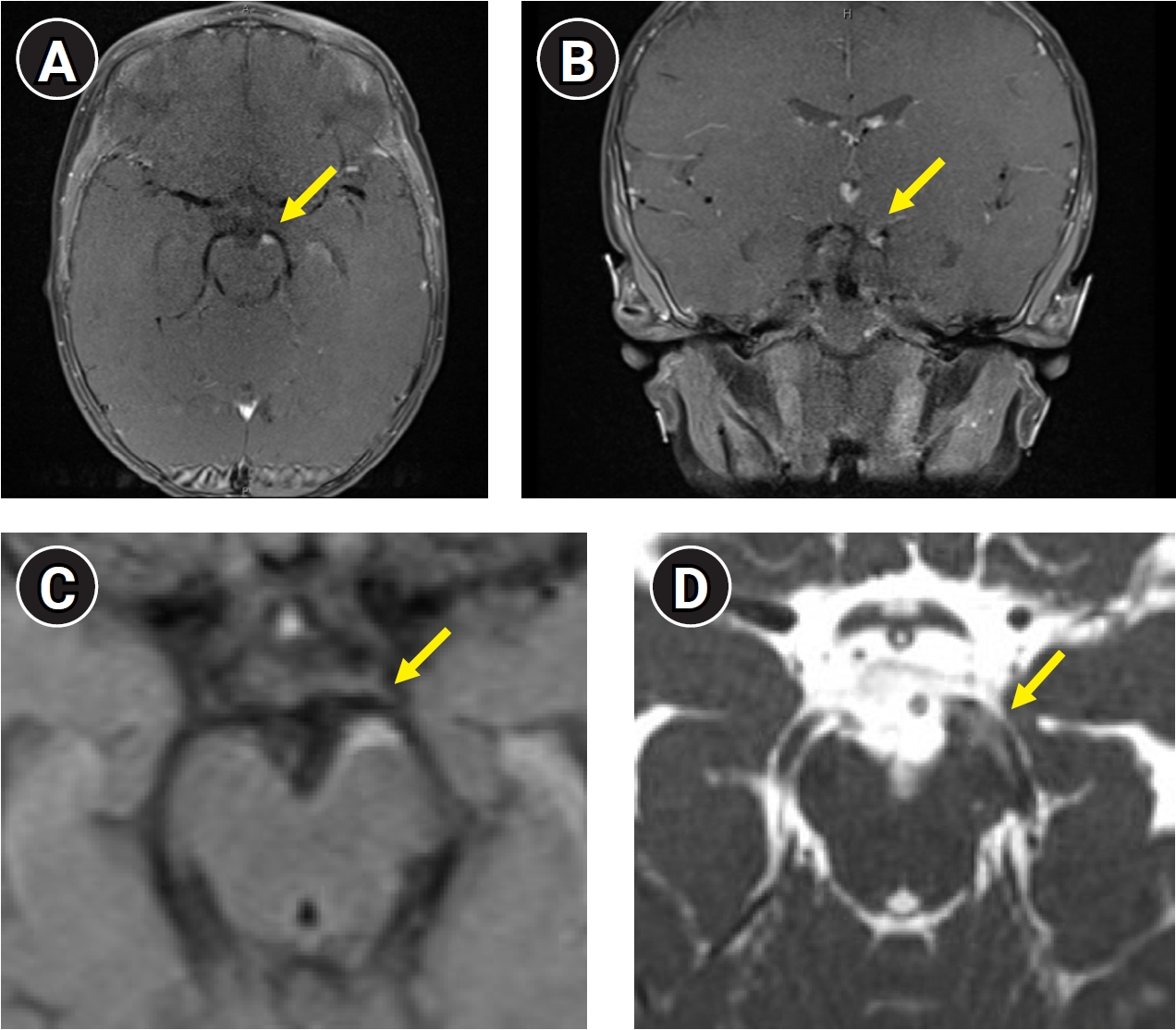
Magnetic resonance imaging of the brain and orbits showing abnormal nodular thickening and enhancement of the proximal cisternal segment of the left oculomotor nerve. (A) Axial postcontrast T1-weighted image showing nodular thickening and enhancement of the left oculomotor nerve (arrow). (B) Coronal postcontrast T1-weighted image showing persistent enhancement and asymmetry of the left oculomotor nerve compared with the contralateral nerve (arrow). (C) Corresponding axial fluid-attenuated inversion recovery sequence showing hyperintensity in the same segment of the left oculomotor nerve (arrow). (D) Corresponding axial constructive interference in steady state sequence further showing abnormal signal in the proximal cisternal segment of the left oculomotor nerve (arrow).
Two weeks after discharge, he was seen in the neurology clinic. His symptoms had improved, although he continued to have mild left eye ptosis and a subtle lag with tracking. By his subsequent follow-up visit 2 months after his initial presentation, his symptoms had completely resolved. A repeat MRI was deferred given his favorable clinical course.
The patient remained asymptomatic until 2 years of age, when he experienced a recurrent episode of isolated left eye ptosis without associated disconjugate gaze. Unlike his initial presentation, his mother denied irritability during this episode. The ptosis resolved spontaneously after 3 weeks, and medical attention was not sought during the episode. One week after symptom resolution, he was hospitalized for intravenous hydration for presumed viral gastroenteritis. Neurologic examination at that time was normal, with resolution of the ptosis and normal extraocular movements, including conjugate gaze and appropriate tracking.
DISCUSSION
The differential diagnosis for acute onset ophthalmoplegia is broad including vascular, infectious, autoimmune and inflammatory etiologies. In order to differentiate RPON from these other entities, clinical evaluations including imaging, serum and cerebrospinal fluid testing should be explored as demonstrated in our patient. The diagnosis of presumed RPON in infants is particularly challenging, as their inability to verbalize migraine-like symptoms necessitates reliance on nonspecific signs such as irritability or crying to infer pain. There are inherent limitations in applying the adult ICHD-3 diagnostic criteria to preverbal infants. Although our patient did not technically meet the diagnostic criteria for RPON due to the inability to accurately assess for a headache, the presence of characteristic MRI findings may provide important supportive evidence in preverbal infants in whom headache symptoms cannot be reliably documented.
The evaluation of an infant presenting with acute ophthalmoplegia requires consideration of a broad range of differentials which include structural lesions, neuromuscular disorders, inflammatory or infectious cranial neuropathies, and congenital cranial dysinnervation disorders. A structural lesion such as a cranial nerve schwannoma was considered. However, our patient’s discrete episodes of ophthalmoplegia with complete interval recovery were less consistent with a structural lesion, which would generally present with progressive or persistent neurologic deficits.6 Additional diagnoses considered include neuromuscular disorders such as myasthenia gravis and Miller Fisher Syndrome (MFS).7 In our patient, the fluctuating nature of his symptoms initially raised concern for myasthenia gravis; however, the corresponding antibodies were negative and cranial nerve enhancement is not a typical characteristic of myasthenia gravis. Although MFS can produce nerve root enhancement, our patient lacked other characteristic features including ataxia and areflexia. Additionally, MFS is classically monophasic whereas RPON causes recurrent attacks over months to years which is consistent with our patient’s episodes at 10 months old and again at 2 years old.8 A congenital cranial dysinnervation disorder may also be considered; however, these disorders typically present with ocular motility abnormalities from birth and have a persistent nonfluctuating course. The associated imaging findings typically reflect developmental anomalies of the cranial nerve rather than focal nerve thickening and enhancement. Our patient’s findings are more suggestive of an acquired process rather than congenital.9
An infectious neuritis is less likely given the absence of fever, leukocytosis, elevated inflammatory markers and his general well appearance. Overall, the leading consideration was a presumed inflammatory oculomotor neuropathy given the recurrent resolving episodes of isolated oculomotor nerve dysfunction in the absence of additional neurologic deficits or systemic inflammatory features. Tolosa–Hunt syndrome is an inflammatory condition that may also present with painful ophthalmoplegia. However, this was considered less likely given its typical localization to the cavernous sinus or superior orbital fissure which may cause inflammation of multiple cranial nerves including the oculomotor, trochlear, abducens, and trigeminal nerves. In contrast, our patient demonstrated isolated oculomotor nerve involvement with enhancement of the cisternal segment of the nerve on MRI and a normal appearing cavernous sinus.10
Although the prognosis for RPON is favorable, several reports have documented recurrent attacks. Lance’s study reports a case of a 9-year-old with three previous episodes at 3, 5, and 12 months old all occurring in the setting of receiving vaccines. Additionally, he discusses a 16-year-old patient with ongoing attacks since 9 months old.11 For our patient, his initial onset was at 10 months old with a recurrent episode at 2 years old. Peck’s study12 also describes a patient with recurrent episodes every 1–2 years beginning at 3 months of age, with episodes becoming less frequent after 9 years of age. Notably, our patient demonstrated the classic finding of cranial nerve III enhancement, whereas previous reports of RPON in patients younger than 1 year described normal MRI studies or the cases pre-dated the widespread use of MRI (Table 1).

Comparison of previous reports discussing cases of recurrent painful ophthalmoplegic neuropathy in patients less than 1 year old
An important limitation of this case is the absence of follow-up MRI to document resolution of the initial nodular thickening and enhancement of the oculomotor nerve, which would have helped to exclude a persistent structural abnormality. Additionally, MRI was not obtained during the recurrent episode of ptosis, limiting confirmation that this episode represented recurrence of the same oculomotor neuropathy. Although the second episode differed phenotypically, without disconjugate gaze or irritability, the recurrence of ipsilateral oculomotor nerve dysfunction followed by complete recovery raises the possibility that it represented a milder recurrence of presumed RPON. However, in the absence of a repeat MRI it cannot be definitively established that both episodes reflected the same underlying pathophysiologic process. Furthermore, because spontaneous recovery has been reported in similar cases, it remains unclear whether the patient's improvement was attributable to corticosteroid therapy or reflected the natural course of the disease. Additional research is needed to establish evidence-based therapeutic recommendations for RPON.
Conclusion
In summary, RPON can have an onset before 1 year of age with accompanying MRI findings of oculomotor nerve enhancement. This report also emphasizes the importance of imaging in establishing the diagnosis, particularly in patients whose headache symptoms cannot be reliably assessed. Because the pathophysiology of RPON remains unclear, no standardized treatment has been established. Systemic steroids have become the most commonly utilized acute intervention and were also applied to our patient’s case. However, given the spontaneous resolution of his symptoms over time, the contribution of corticosteroid treatment to his clinical improvement could not be determined, particularly because RPON commonly exhibits spontaneous recovery. Further studies are needed to evaluate the pathophysiology of this condition to help guide future treatments.
Notes
AVAILABILITY OF DATA AND MATERIAL
The datasets are not publicly available because they contain confidential information but are available from the corresponding author upon reasonable request.
AUTHOR CONTRIBUTIONS
Conceptualization: KMY, RHF; Writing–original draft: KMY; Writing–review & editing: KMY, RHF.
CONFLICT OF INTEREST
No potential conflict of interest relevant to this article was reported.
FUNDING STATEMENT
None.
ACKNOWLEDGMENTS
None.